|
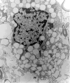
Fig 2. Electron micrograph of a neural cell with extensive cytoplasmic vacuolization. A small myelinated axon can also be seen. The tissue was obtained from the second brain biopsy. Magnification X 11,700
|
|
Fig 2. Electron micrograph of a neural cell with extensive cytoplasmic vacuolization. A small myelinated axon can also be seen. The tissue was obtained from the second brain biopsy. Magnification X 11,700
|