|
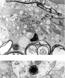
Fig 4. Electron micrograph of a cell showing vaccuolization, mitochondrial disruption and a prominent inclusion containing membranous and other darkly staining components. Blebbing and degeneration of portions of the myelin sheaths can be seen. Axoplasmic material is lacking from some of the myelin sheaths. An enveloped particle, consistent with a virus, is seen within a vacuole of the cell, and is shown at X 183,000 in the lower frame. Mag. X 17,200
|