|
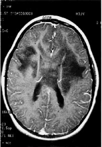
Fig 5. T1 weighted MRI of the brain. The corresponding T2 weighted image showed heightened intensity of the affected areas. There was no post-gadolinium enhancement. The affected regions included both frontal lobes, and the anterior body of the corpus callosum, seen as the curved body crossing the midline.
|