|
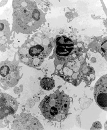
Fig 6. Electron microscopy of Ficoll-Paque separated mononuclear cells obtained from a blood sample collected in an ACD vacutainer tube. Note the vacuolization and signs of cellular degeneration. Mag. X 5,900
|
|
Fig 6. Electron microscopy of Ficoll-Paque separated mononuclear cells obtained from a blood sample collected in an ACD vacutainer tube. Note the vacuolization and signs of cellular degeneration. Mag. X 5,900
|