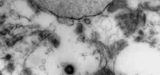
 Fig. 13: Electron micrograph of a degenerate, vacuolated cell from the brain biopsy from patient 3. Note the thickened patches along the nuclear membrane and the cytoplasmic collections of granular and microvesicular components. Several descrete particles can also be seen (X 50,600). |