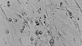
 Fig. 14: Phase contrast of the early CPE seen in MRHF fibroblast cultures inoculated with material obtained from the brain biopsy of patient 3. The normally spindle shaped fibroblasts become round and show progressive swelling with subsequent evidence of syncytia formation (X 250). |