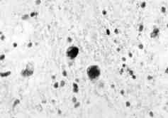
 Fig. 3: The region of the section from the stereotactic biopsy shows two enlarged, vesiculated cells with grossly distorted nuclei. The cells stained strongly with PAS (X 400). |
|
Fig. 3: The region of the section from the stereotactic biopsy shows two enlarged, vesiculated cells with grossly distorted nuclei. The cells stained strongly with PAS (X 400). |