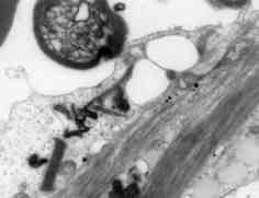
 Fig. 4: Electron micrograph from patient 1 showing lipid vacuoles within a glial cell. Several small densely staining spherical structures are seen within the cell and have a viral-like appearance. A myelin sheath is seen adjacent to the glial cell (X 28,750). |