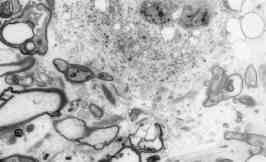
 Fig. 5: Electron micrograph from patient 1 showing a region of accumulation of irregularly sized particles within a cell with disrupted mitochondria. Several of the myelin sheathes do not contain axons and show evidence suggestive of disintegration (X 7,820). |