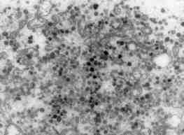
 Fig. 6: Electron micrograph of an additional region of the biopsy from patient 1 showing more details of the irregularly sized particles (X 28,750). |
|
Fig. 6: Electron micrograph of an additional region of the biopsy from patient 1 showing more details of the irregularly sized particles (X 28,750). |