 Return to article
|
|
|
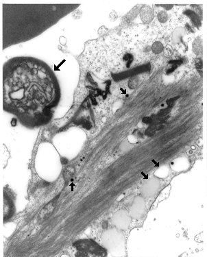
Electron micrograph of a vacuolated glial cell seen in a stereotactic brain biopsy from a patient with a stealth virus encephalopathy. Small, dark, viral-like particles can be seen. Note the adjacent empty and lipid-filled vacuoles in the lower right side of the cell. The round body in the upper left is an axon surrounded by a myelin sheath.
Return to article
|